MRI

Magnetic resonance imaging (MRI) is a non-invasive diagnostic procedure that uses a magnetic field, radio waves, and a computer to produce detailed pictures of internal structures of the head and body. MRI excels at seeing soft tissues, and can provide clearer differentiation between diseased and normal tissue than other imaging methods. Because MRI does not use radiation (x-rays), it poses almost no risk to the average patient.

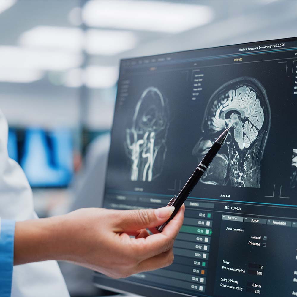
Radiologist looks at patient MRI brain scan

MRI

Magnetic resonance imaging (MRI) is a non-invasive diagnostic procedure that uses a magnetic field, radio waves, and a computer to produce detailed pictures of internal structures of the head and body. MRI excels at seeing soft tissues, and can provide clearer differentiation between diseased and normal tissue than other imaging methods. Because MRI does not use radiation (x-rays), it poses almost no risk to the average patient.

MRI is commonly used to diagnose problems in the brain and spinal cord. MRI is also used to examine organs of the abdomen and pelvis, as well as the joints and other musculoskeletal structures. MR Angiography (MRA) specifically focuses on examination of the circulatory system, to help diagnose abnormalities of the heart and blood vessels, congenital abnormalities, arterial disease, vessel obstruction, injury to arteries caused by trauma, and more.

Our radiologists perform MRIs using a variety of equipment.

In association with Merrimack Valley Health Services, Lawrence General Hospital offers a 3T outpatient facility on Route 125 in North Andover and a 1.0T high-field open magnet of the LGH campus in Lawrence.

3 Tesla MRI (3T MRI) is presently the strongest clinical MRI in use, providing faster and clearer imaging than previous MRI technologies. 3T MRI is particularly useful when the details are crucial to diagnosis, such as imaging small bones, breast tissue, musculoskeletal,  neurological, and vascular structures. However, just because 3T is the strongest, it does not mean it is the best choice for every situation. Our radiologists will work with you to determine the best option for you.

Open MRI scanners are open on all the sides, unlike the tunnel structure of a typical MRI scanner. Because open MRIs tend to use lower magnet strength, some imaging cannot be performed with these scanners. However, when appropriate, open MRIs can be beneficial for patients who are claustrophobic, who require acute care supervision, or who are too large for traditional MRI scanners.

Preparation

Preparation instructions vary from one MRI procedure to the next — some procedures may require you not to eat for a few hours or may require use of an intravenous contrast dye. We will provide you with instructions specific to your procedure beforehand.

Metal and electronics may cause a safety hazard or interfere with the magnetic field during an MRI and are not allowed in the room. These items include body piercings and other jewelry, hair pins, eyeglasses, dentures, pens, hearing aids, and purses, shoes, and clothing that contains metal, such as pants with zippers or bras with metal underwire. Notify your physician if your body contains any bullets or shrapnel, or if you have any medical implants or devices, including cochlear implants, pacemakers, artificial joints or valves, metal pins or screws, and aneurysm clips. It may not be safe for you to have an MRI, and your physical may opt for another type of imaging.

Always inform your physician of recent illnesses, chronic medical conditions, and whether there’s a chance you may be pregnant before any procedure is performed. Wear comfortable clothing; you may be asked to change into a gown.

Copyright © 2025 - L&M Radiology | Website by Cold Spring